Prenatal ultrasound | Prenatale echo

2025 - 70x70 cm
- Enlarged nuchal translucency
- Enlarged cisterna magna
- Mild ventriculomegaly
- Choroid plexus cysts
- Absent nasal bone
- Tricicuspid regurgitation
- Echogenic focus in heart
- Renal pyelectasis
- Echogenic bowel
- Short femur
- Single umbilical artery
- Amniocentesis
- Chorionic villus sampling
- Placenta
- Turbulant sea
- Stork incoming
- Some clouds
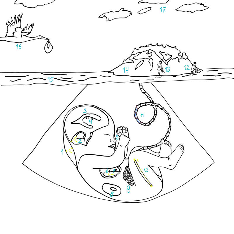
Prenatal soft markers
During pregnancy, ultrasound examinations of the baby can be performed at various moments. Fortunately, no abnormalities are seen in most babies.
Sometimes, however, so-called soft markers are detected. These are subtle ultrasound findings that are often temporary and may disappear over time. On their own, they are usually harmless, but in some cases they can indicate that there may be more going on, such as a chromosomal abnormality in the baby. In this painting, I have depicted 11 of these soft markers in the baby. When only one soft marker is seen, the likelihood of a genetic abnormality is generally small. However, the more soft markers that are detected, the greater the chance that there may be an underlying genetic condition.
Many pregnant women and their partners are already looking forward to the arrival of their child and are preparing for this new chapter (in the Netherlands the stork is on its way). When something unusual is seen on the ultrasound, it understandably causes considerable anxiety for the parents. In the painting, this is symbolized by the turbulent sea and clouds in the sky.
Depending on the number and type of soft markers, there may be a reason to perform invasive prenatal diagnostic testing. This can be done through chorionic villus sampling (CVS) or amniocentesis. During CVS, a small sample of placental tissue is taken. In the painting, this is represented by a gynecologist holding a small scoop. During amniocentesis, a needle is used to withdraw a small amount of amniotic fluid. This is depicted in the painting as a gynecologist holding a small bucket.

Prenatale soft markers
- Verdikte nekplooi
- Vergrote cysterna magna
- Milde ventriculomegalie
- Plexus choroïdeus cysten
- Afwezig neusbeen
- Tricicuspidalis regurgitatie
- Echogene focus in hart
- Pyelectasis nieren
- Echogene darm
- Kort femur
- Enkele navelstrengarterie
- Vruchtwaterpunctie
- Vlokkentest
- Placenta
- Turbulente zee
- Ooievaar in aantocht
- Een paar wolken
Tijdens een zwangerschap kan op verschillende momenten een echo van de baby worden verricht. Gelukkig worden er bij de meeste baby’s geen afwijkingen gezien.
Soms worden echter zogenaamde soft markers gezien. Dit zijn subtiele echo bevindingen die vaak tijdelijk zijn en weer verdwijnen. Op zichzelf zijn ze meestal onschuldig maar soms kunnen ze erop wijzen dat er meer aan de hand is, zoals bijvoorbeeld een chromosoomafwijking bij de baby. Op het schilderij heb ik 11 van dit soort soft markers bij de baby geschilderd. Als er maar één soft marker wordt gezien, is de kans op een genetische afwijking bij de baby meestal klein. Maar hoe meer soft markers er bij de baby worden gezien hoe groter de kans dat er sprake kan zijn van een onderliggende genetische afwijking.
Veel zwangeren en hun partners zijn zich al aan het verheugen op de komst van een kind en de voorbereidingen hiervoor aan het treffen (de ooievaar komt eraan). Op het moment dat er op de echo iets bijzonders bij het kind wordt gezien geeft dit vanzelfsprekend veel onrust bij de ouders. Dit wordt op het schilderij gesymboliseerd door de woelige zee en wolkjes in de lucht.
Afhankelijk van het aantal en het type soft markers kan er een reden zijn om invasieve prenatale diagnostiek te verrichten. Dit kan gedaan worden met een vlokkentest of een vruchtwaterpunctie. Bij de vlokkentest wordt er een stukje van de placenta (moederkoek) weggenomen. Op het schilderij is dit verbeeld door een gynaecoloog met een schepje. Bij een vruchtwaterpunctie wordt met een naald wat vruchtwater opgezogen. Dit is in het schilderij weergegeven door een gynaecoloog met een emmertje.